
ALL MATERIAL COPYRIGHT KEVIN SCOTT 2011. LINKS TO THIS SITE ARE WELCOME BUT DO NOT COPY MATERIAL FROM THIS SITE TO ANY OTHER WEBPAGE.


If you find this site useful, please support it by making a donation of $1 to help maintain and develop it. Click on the PAYPAL DONATE button to do this safely. But there is no obligation - please avail yourself of the information and facilities of the site at no charge.
Chemical Microscopy is the technique of using the optical microscope to observe chemical reactions taking place on microscope slides usually for analytical purposes. The conduct of qualitative inorganic Chemical analysis under the microscope was pioneered in the first half of the 20th century. A major reference work on the subject was published in the 1950s entitled "A Handbook of Chemical Microscopy" which was a comprehensive description of both the techniques involved and a catalogue of a large number of analytical tests which could be performed on a microscope slide. Much of what appears in these pages is derived from this work.
Carrying out qualitative inorganic analysis on a microscope slide presents considerable advantages over the conventional technique. By optimising the conditions of any particular reaction, it is possible to arrange that crystalline precipitates which form do so with a readily observable crystal structure. This provides much more information than would be provided by the reaction viewed with the naked eye. Additionally, techniques such as polarimetry, and the use of fluorescence can provide significantly useful information on the substances being observed.
It has to be said that Chemical Microscopy as an analytical tool has been largely superseded by the vast array of analytical instrumental techniques which are now available. It was overtaken so rapidly in analytical laboratories that it really scarcely enjoyed a heyday and was consigned to history almost at its conception. However, it has long been a conviction of the author of these present pages, that optical microscopy has generally been seriously underused in the chemical laboratory. It is therefore proposed to revisit the technique of chemical microscopy to give it, so to speak, its day in court, to examine its virtues and explore its possibilities. The modern developments in photo micrography encourage this ambition. When the handbook of chemical microscopy was published all the visual information has to be transmitted to the reader via black-and-white sketches. A simple electronic camera attached to a microscope can provide high resolution graphics which communicate much better than the sketches. The quality of information potentially available from Chemical microscopy is thus now greatly enhanced, as it is hoped these pages will illustrate.
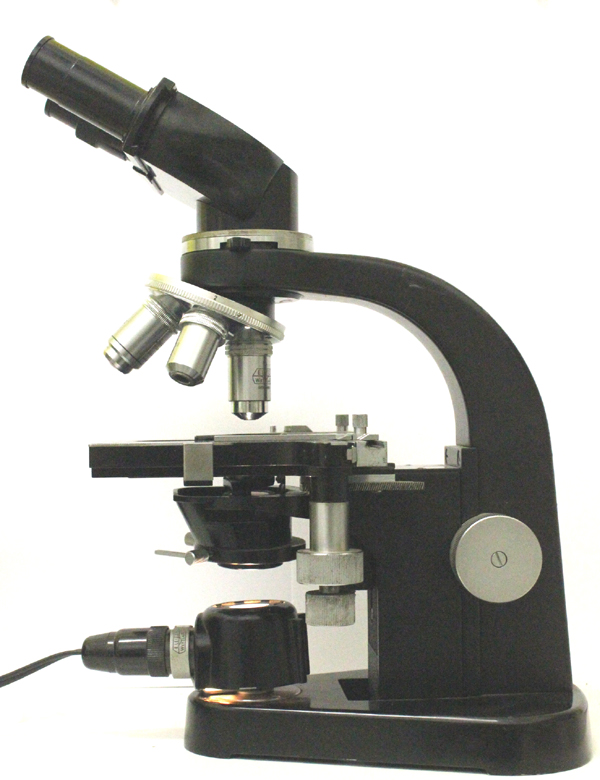
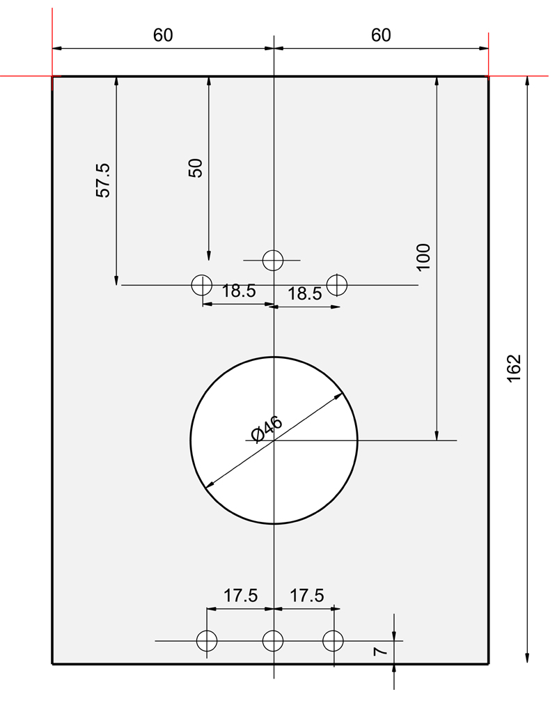
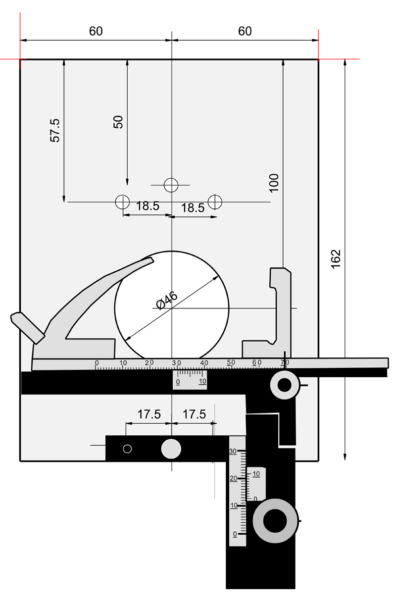
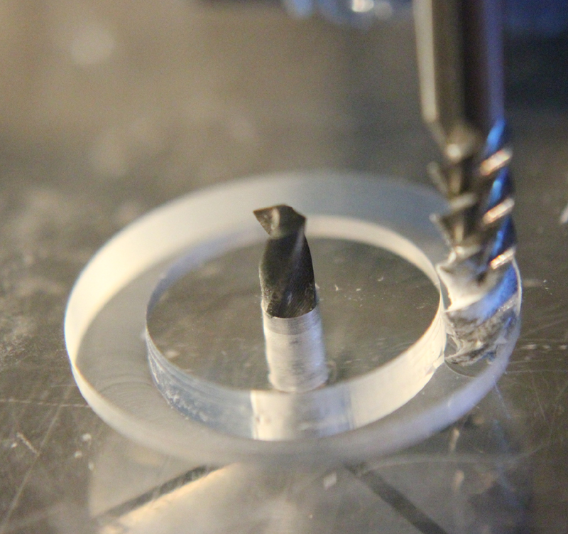
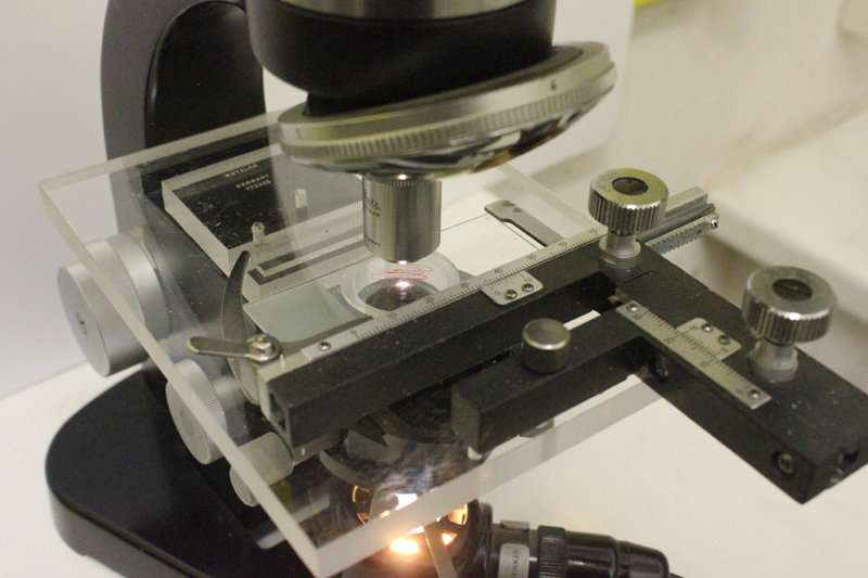
The microscope selected for use in chemical microscopy in this case was a Leitz Wetzlar binocular model as shown in the photograph above. The chemical laboratory is, of course, a harsh environment for a precision optical instrument and some modifications are desirable to minimise the corrosion of the microscope which could otherwise occur. The principle such modification was the replacement of the stage of the microscope with one machined from acrylic sheet. The regular metal stage was removed by dismantling the microscope frame. The removal of four hex socket cap screws from the underside of the base enabled the latter's removal and, after removal of the condenser, allowed access to the four screws which secured the stage to the instrument. Once this had been removed, and acrylic replacement could be made according to the diagrams below.
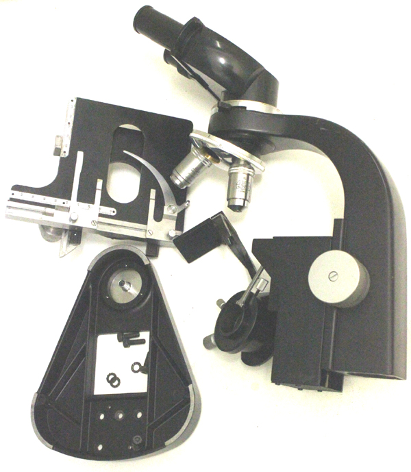
The original mechanical stage was not used, but another, less expensive generic version substituted, which could be easily replaced if it suffered damage or corrosion. An aperture was machined in a position collinear with the optical path which could be fitted with a replaceable acrylic disc if required.
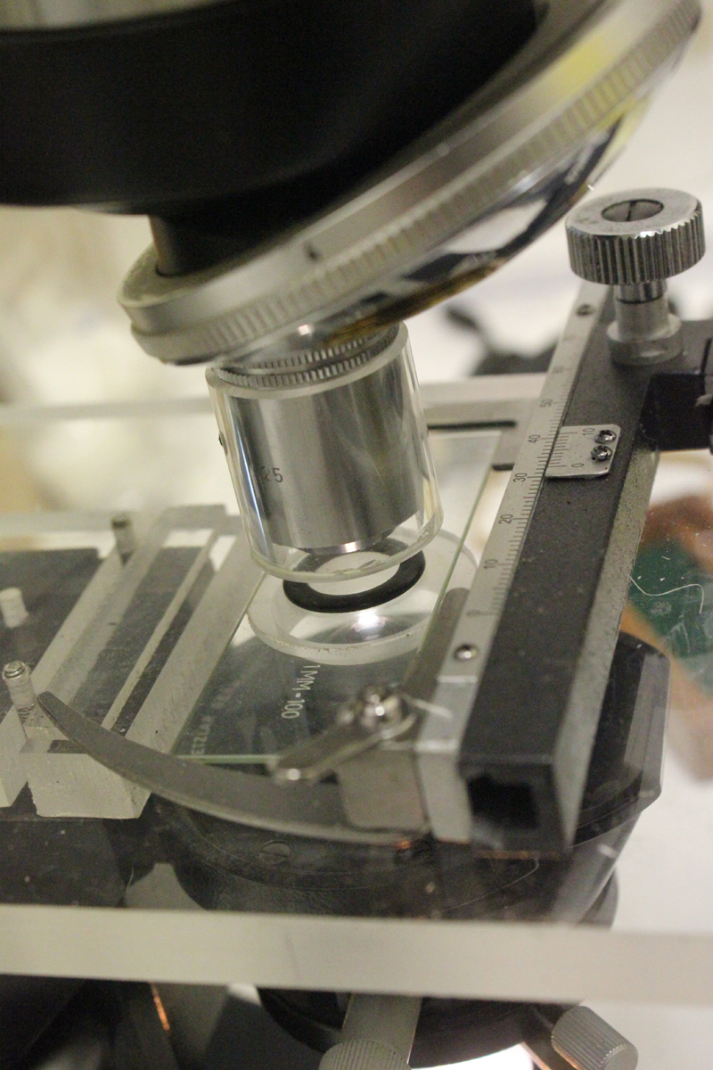
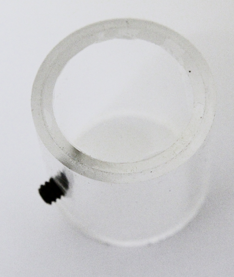
One further modification of the microscope was considered desirable. To prevent corrosion of the objective lens, a close fitting acrylic tube was machined and fitted with a 22 mm diameter glass coverslip at one end and a 6-32 setscrew was provided to enable the constructed objective guard to be secured to the objective. This is illustrated to the right.
Photomicrography with reasonably high resolution can now be readily achieved using simple optics and USB cameras. A DCM900 Scopetek camera was fitted with an optical coupling giving a magnification of 0.4x and providing a 23.2mm insertion tube to replace a standard eyepiece. The purpose of the 0.4x interposing optics between the eyepiece socket on the microscope and the camera was to match the field of view obtained using a standard eyepiece to that obtained by the camera. The camera is connected to a laptop computer running Windows XP via a USB cable and a TWAIN driver installed on the computer. This allowed image processing software such as Adobe photoshop or or serif photoplus to acquire images from the microscope.
The attainable resolution was estimated by gathering images from a stage micrometer having divisions 10 µm apart. These were clearly resolved and the maximum resolution obtainable was estimated at about 2 μm. This compares with the theoretical maximum resolution obtained with the particular objective used of about 1 μm